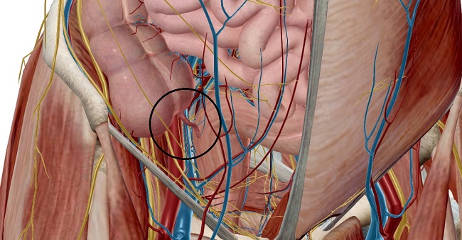
The vermiform appendix is a narrow, blind-ending intestinal diverticulum arising from the posteromedial aspect of the caecum, 2-3cm inferolateral to the ileocecal valve.
Structure
It is usually 6-10 cm long in the adult with approximate diameter of the lumen about 6mm. It consists of base, lumen and tip. The lumen is wide in infants and obliterated in elderly people. The luminal orifice is sometimes guarded by semi-circular fold of mucous membrane known as Gerlach’s valve. The appendix has a continuous outer layer of longitudinal muscle formed by the coalescence of the three taeniae coli.

It has triangular fat containing mesentery running between the terminal ileum and appendix known as mesoappendix. A small fold of peritoneum also runs between the terminal ileum and anterior layer of mesoappendix known as “bloodless fold of Treves”. Appendix consists of masses of submucosal lymphoid tissue, hence called “Abdominal tonsil”.
Embryology
The vermiform appendix arises from the midgut. The caecal diverticulum, precursor of the vermiform appendix appears at 6th week and appendix is visible by 8th week. The appendix undergoes medial rotation along with midgut and descends into the right lower abdomen at 10th week. The mucosa develops lymphoid tissue by 14-15 weeks.
Position of Appendix
The appendix lies in the right iliac fossa. Although the base of the appendix is fixed, surfacely marked by McBurney’s point on spino-umbilical line (two-thirds of the way along a line between umbilicus and anterior superior iliac spine), the tip can be in any direction giving the variable positions. With no fixed position, it is known to be the organ of the body with no fixed anatomical position. The anatomical position of the appendix also determines the symptoms and site of muscular spasms and tenderness when the appendix is inflamed.
- Retrocolic or retrocaecal: most common (65%), behind the caecum or lower ascending colon.
- Pelvic: descends over the pelvic bream (30%).
- Splenic: towards the spleen as pre-ileal or post-ileal (anterior or posterior to the terminal ileum respectively.
- Promontoric: towards the sacral promontory.
- Paracolic: upwards and towards the right.
- Subcaecal or mid-inguinal: below the caecum and towards the inguinal ligament.
Vascular supply and Lymphatic drainage
Arterial supply
The main artery is appendicular artery, branch of inferior division of ileocolic artery from superior mesenteric artery. Next is accessory appendicular artery, branch of posterior cecal artery.
Venous drainage
The appendicular vein draining the appendix joins the cecal vein to become ileocolic vein, which in turn joins the portal vein.
Lymphatic drainage
Lymphatic drainage is to lymph nodes of mesoappendix and ileocolic lymph nodes which drains into superior mesenteric lymph nodes.
Nerve supply
The autonomic innervation of the appendix arises from the superior mesenteric plexus. Sympathetic supply originate from the lower thoracic part (T9 and T10) of the spinal cord while parasympathetic supply is from the vagus. Affarent fibres from the appendix accompany the sympathetic nerves to the T10 segment of the spinal cord.
Functional Anatomy
Although thought to be vestigial organ, appendix serves the following functions:
- It acts as a reservoir for gut flora, so beneficial bacteria repopulate in the digestive system to replace the bacteria cleared by excessive bouts of diarrhea.
- It is an important component of the mucosal immune system, mainly B cell-mediated immune response.
Clinical Anatomy
- Appendicitis is the inflammation of the appendix characterized by the abdominal pain around umbilicus and then tenderness in McBurney’s point, nausea, anorexia, vomiting etc. Appendectomy, surgical removal of the appendix is usually necessary. It is generally uncommon in two extremes of life, infancy and elderly.
- In appendicitis, extension of hip causes pain due to the spasm of psoas major in retrocaecal position and flexion and medial rotation causes pain due to spasm of obturator internus in pelvic position.
- In unusual cases of malrotation of intestine or failure of descend of caecum, the appendix lies in the right hypochondriac region. So, pain localizing in this level may mimic signs and symptoms of acute cholecystitis. This type of appendix is known as sub-hepatic appendix.
.
.
References
- Gray’s Anatomy,The Anatomical Basis Of Clinical Practice, forty-first edition.
- Moore’s Clinically Oriented Anatomy, seventh edition.
- Vishram Singh’s Clinical and Surgical Anatomy, second edition.
- BD Chaurasia’s Human Anatomy, seventh edition.
- The Appendix, Wikipedia.
- Research Journal on Appendix by Bonnie D. Hodge, Sarang Kashyap, Arshia Khorasani-Zadeh.
- Langman’s Medical Embryology, twelfth edition.

